Why Diagnostic Cast Dental Still Matter in the Digital Age?
In an era where digital intraoral scanning and 3D imaging are advancing rapidly, the diagnostic cast remains a cornerstone in clinical dentistry. Often underestimated, this simple yet powerful tool is indispensable in diagnosis, treatment planning, and patient communication across multiple dental disciplines.
A diagnostic cast sometimes called a study model is a three dimensional replica of the teeth, surrounding tissues, and oral structures, fabricated from impressions taken during the initial stages of dental examination. Despite the digital revolution, many procedures still depend on this tactile, physical model for precision planning.

What Is a Diagnostic Cast in Dentistry?
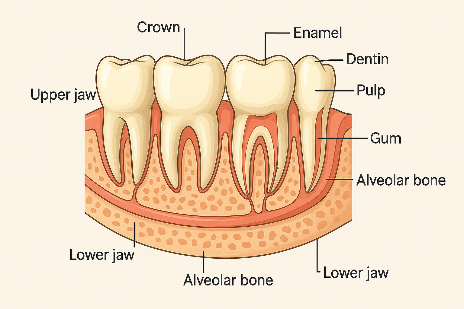
A diagnostic cast dental model is a highly accurate, gypsum-based replica of a patient’s oral cavity, created to reflect their teeth structure, gum tissues (gingiva), and bite alignment (occlusion). This physical model is essential in clinical dental assessments, typically produced prior to starting dental treatment. It helps dental professionals analyze complex dental conditions beyond what is visible during a standard intraoral examination.
This three dimensional dental model serves as a permanent dental record of the patient’s oral health status and is crucial in comprehensive treatment planning. The diagnostic cast provides clarity on several key clinical aspects, including:
- Malocclusion and bite relationship – It assists in diagnosing overbites, underbites, and other occlusal discrepancies, improving orthodontic evaluations.
- Tooth alignment and rotations – Highlights dental crowding, spacing issues, and tooth angulation, supporting orthodontic planning.
- Tooth wear, fractures, or spacing – Detects enamel erosion, chipped teeth, and abnormal gaps that may require restorative procedures.
- Periodontal conditions – Reveals gum recession, gingival contours, and potential signs of periodontal disease, aiding in periodontal diagnosis.
By offering a tangible, static representation of the mouth, the diagnostic dental cast enhances the accuracy of oral health evaluations, supporting prosthodontic, restorative, and orthodontic treatments.
How Are Diagnostic Dental Casts Made?
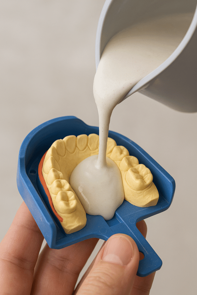
The creation of a diagnostic dental cast involves two critical steps: dental impression taking and gypsum model pouring. These steps ensure the production of a highly accurate, three dimensional replica of a patient’s oral structures, which is essential for dental diagnosis and treatment planning.
Impression Taking
The first stage involves capturing an exact impression of the dental arches (both maxillary and mandibular). This is done using high precision impression materials such as:
- Alginate (irreversible hydrocolloid) – Widely used for preliminary impressions due to its quick setting time and ease of manipulation.
- Polyvinyl siloxane (PVS) – An elastomeric impression material known for its dimensional accuracy and stability over time.
- Polyether – Offers excellent wetting ability, flow characteristics, and precise reproduction of oral tissues.
These materials accurately capture the tooth anatomy, gingival contours, and overall oral cavity structure required for a reliable dental model.
Model Pouring
After the impression is taken, it is filled promptly with dental stone to create the physical model. The commonly used gypsum materials include:
- Type III gypsum (Model Stone) – Used for standard diagnostic casts requiring moderate strength and surface detail.
- Type IV gypsum (High-Strength Die Stone) – Preferred for more complex cases needing greater hardness, fine detail, and dimensional stability.
Once the gypsum material sets, the cast is trimmed, smoothed, and, when necessary, mounted on a dental articulator to simulate the patient’s bite relationship and occlusal function. This step by step process results in a highly detailed diagnostic cast, which acts as a reliable visual aid for clinical examination, prosthodontic planning, and orthodontic evaluation.
Clinical Applications of Diagnostic Casts in Modern Dentistry
Diagnostic dental casts are essential tools in various disciplines of modern dentistry. They offer a three dimensional representation of the patient’s oral anatomy, allowing for both comprehensive diagnostic evaluation and treatment planning. Their accuracy supports informed decision making across prosthodontics, orthodontics, and restorative dentistry.

In Prosthodontics
In prosthodontic treatment, diagnostic casts assist in:
- Evaluating edentulous ridges for denture base design and support.
- Measuring vertical dimension of occlusion to maintain facial harmony and bite function.
- Analyzing the occlusal plane for proper alignment of artificial teeth.
These models are integral to fabricating complete dentures, ceramic crowns, and fixed dental bridges that offer optimal fit, retention, and functionality. They also support implant planning in edentulous cases.
In Orthodontics
In the field of orthodontic diagnostics, dental casts provide valuable data for:
- Space analysis to assess arch length discrepancies.
- Evaluating overjet and overbite to determine the extent of malocclusion.
- Measuring tooth crowding and rotation patterns.
- Simulating tooth movement for advanced orthodontic treatment planning.
In Restorative Dentistry
In restorative and cosmetic dentistry, diagnostic casts are used to:
- Design full mouth occlusal rehabilitation plans for patients with worn dentition.
- Plan and visualize inlays, analysis, and indirect restorations for precise fit and esthetics.
- Develop esthetic mock ups for smile design previews and patient consultations.
These applications enhance functional restoration, support minimally invasive procedures, and improve patient communication during aesthetic treatment planning.
Difference Between a Preliminary Cast and a Diagnostic Cast
Though similar in appearance, a preliminary cast and a diagnostic cast differ in precision and clinical value.
| Feature | Preliminary Cast | Diagnostic Cast |
| Accuracy Level | Moderate | High precision |
| Purpose | Initial evaluation or record-keeping | Detailed treatment and prosthetic planning |
| Mounting | Rarely mounted | Often mounted on articulators |
| Usage | Rough visualization | Functional simulation and analysis |
According to the American College of Prosthodontists, diagnostic casts should be incorporated in all fixed and removable prosthodontic cases to support predictable clinical outcomes.
Why Diagnostic Casts Are Crucial for Long Term Dental Success?
An accurately made cast can prevent restorative failures, reduce post-op complications, and ensure that prosthetic or orthodontic appliances are customized for the patient’s functional anatomy.
Poor quality or skipped casts can lead to:
- Misaligned restorations
- Improper occlusion
- Gingival irritation or trauma
- Increased cost due to adjustments or remakes
Diagnostic Casts in Patient Communication and Education
One of the most valuable yet often overlooked functions of a diagnostic dental cast is its role in enhancing patient communication and oral health education. Unlike intraoral scanners or digital radiographs, which can be difficult for non clinicians to interpret, a physical dental model provides a clear, three-dimensional view of the patient’s oral condition. This visual aid in dentistry helps bridge the gap between clinical diagnosis and patient understanding.
Key Benefits
- Tangible Visualization: Dentists can physically demonstrate tooth wear, dental fractures, malocclusion, and spacing abnormalities using the cast. This hands on approach simplifies dental condition explanation and enhances patient awareness.
- Before and After Simulations: Diagnostic casts can show projected treatment outcomes, such as the results of orthodontic treatment, restorative procedures, or cosmetic enhancements. Patients can clearly visualize anticipated changes in their oral structure.
- Improved Case Acceptance: When patients fully comprehend their oral health condition and the proposed treatment plan, they are more likely to approve the intervention. This approach not only increases treatment acceptance but also helps reduce dental anxiety by making complex procedures more approachable.
Conclusion
A diagnostic cast is a crucial foundation in dentistry, ensuring precision, predictability, and patient-centered care. Despite advances in digital dentistry, 3D scanning, and CAD/CAM technology, physical models made from Type III or IV dental stone remain indispensable. These gypsum-based dental replicas provide detailed, tactile insight into malocclusion, occlusal dynamics, and restoration planning, improving accuracy for crowns, bridges, and implants.
Diagnostic casts enhance communication with dental laboratories, reduce clinical errors, and boost patient understanding and treatment acceptance. Supported by research, they decrease chair time and improve prosthetic fit. Essential in dental education, they help students learn anatomy and case complexities. Serving as permanent medico-legal records, they document oral health over time. Integrating physical models with digital workflows allows modern dentistry to deliver comprehensive, precise, and effective care.
Frequently Asked Questions
Q1: What is the main purpose of a diagnostic dental cast?
It is primarily used to assess a patient’s oral condition in a 3D format, helping in accurate diagnosis, treatment planning, and laboratory communication.
Q2: Can diagnostic casts be used for surgical guides?
Yes, especially when planning implant placements, diagnostic models can be used to fabricate surgical stents or guides with proper angulation.
Q3: Are digital models replacing physical diagnostic casts?
While digital dentistry is growing, physical models still offer superior tactile feedback, articulator simulation, and realistic visualizations that are indispensable in complex cases.
Q4: What materials are best for high quality diagnostic casts?
Type IV gypsum stone offers superior strength, low expansion, and detail reproduction, making it ideal for prosthetic and restorative diagnostics.

 Medically reviewed by